


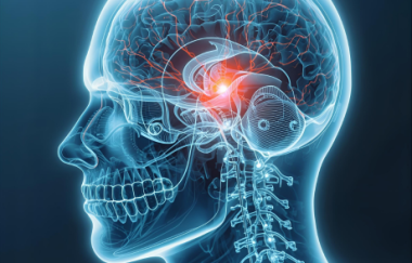
Diffusion-weighted magnetic resonance imaging (DWI) and 1H magnetic resonance spectroscopy (MRS) complement each other in the evaluation of temporal lobe epilepsy. Following surgery for epilepsy, DWI lateralized to the operated side more frequently while 1H MRS was a better predictor of postoperative seizure control, report investigators from the Departments of Diagnostic Radiology and Neurology at the Mayo Clinic in Rochester, Minnesota, United States.
The investigators used DWI and 1H MRS to study 40 temporal lobe epilepsy patients who underwent surgery and 20 normal subjects.
They obtained N-acetylaspartate/creatine ratios in the medial parietal and temporal lobe and also apparent diffusion coefficients of the hippocampal and temporal stem. Results showed that temporal lobe N-acetylaspartate/creatine lateralized to the operated temporal lobe in 45 percent of patients, hippocampal apparent diffusion coefficients in 80 percent of patients and temporal stem apparent diffusion coefficient in 65 percent of patients.


תגובות רוצה להצטרף לדיון?
יש להתחבר כדי להגיב.
התחבראין תגובות עדיין. היה הראשון להגיב!