

כתבות בנושאים דומים

עליה של עשרות אחוזים בשיעורי התמותה על-רקע היריון בשנים האחרונות בארצות הברית (JAMA Netw Open)
בארצות הברית דווח על שיעורי התמותה על-רקע היריון הגבוהים ביותר מבין המדינות בעלות הכנסה-גבוהה, עם למעלה מ-6,000 מקרי תמותה מדווחים בין 2018 עד 2022, כך על-פי נתונים שפורסמו בכתב העת JAMA Network Open. מניתוח הנתונים עלה כי בילידות אלסקה ובאמריקאיות ממוצא אינדיאני תועדו שיעורי התמותה הגבוהים ביותר. מחקר החתך התבסס על נתונים ארציים של המרכז […]

אימוני אינטרוול בעצימות גבוהה מפחיתים סיכון להידרדרות תפקוד כלייתי בקשישים (J Am Society Nephrol)
אימוני אינטרוול בעצימות גבוהה (High-Intensity Interval Training, או HIIT) תחת השגחה לאורך חמש שנים עשויים להפחית את הסיכון להידרדרות מהירה בקצב הפינוי הגלומרולארי המשוער בקשישים בגילאי 70-77 שנים, כך עולה מנתונים חדשים שפורסמו בכתב העת Journal of the American Society of Nephrology. ברקע למחקר מסבירים החוקרים כי למרות היתרונות הבריאותיים הרבים של פעילות גופנית, אין […]

האם המהפכה הטיפולית בהשמנת יתר תפרוץ את חומת הכלכלה? פרופ' רז ופרופ' שטרן
לפניכם הרצאה ודיון שהתקיים בין פרופ’ איתמר רז ופרופ’ נפתלי שטרן בנושא השפעות החידושים הטיפוליים בתחום ההשמנה.
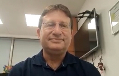
בעקבות המלצות ועדת הסל - ראיון מיוחד עם פרופ' נדב דווידוביץ
על רקע פרסום המלצות ועדת הסל ביקשנו לבדוק עם פרופ’ נדב דווידוביץ, חבר בוועדת הסל, כמה סוגיות שקשורות למתודולוגיה ולאופן קבלת ההחלטות בוועדה.

מאזן עלות-תועלת של הטיפול ב-Dapagliflozin במחלת כליות כרונית (Clin J Am Soc Nephrol)
אנליזה חדשה של מחקר הCKD-DAPA מראה שהטיפול ב-dapagliflozin (פורסיגה) במחלת כליות כרונית הינו משתלם מבחינת מאזן עלות-תועלת בבריטניה, גרמניה וספרד. ברקע למחקר מסבירים החוקרים כי מחלת כליות כרונית (Chronic Kidney Disease CKD) מהווה נטל משמעותי על המטופלים ועל שירותי הבריאות, בעיקר בהגעה לאי ספיקת כליות סופנית כשהמטופלים נדרשים לטיפול כלייתי חליפי. מחקר ה-DAPA-CKD הראה שהטיפול ב-dapagliflozin, […]

איך זה עבר בשקט ומתחת לרדאר? עלות ההשמנה בישראל - 20 מיליארד ש''ח לשנה (הודעת החברה להשמנה)
כבר לפני יותר משנה הושלם דו”ח מקיף, המנתח את השלכות מחלת ההשמנה על חוסן מדינת ישראל, עם השוואות לעולם ותכנית ברורה למדיניות בריאות מסודרת ולרגולציה מותאמת. נחשף היופ, בתום הכנס השנתי של החברה הישראלית לחקר וטיפול בהשמנת יתר של הר”י ד”ר דרור דיקר (בתמונה) : “נהוג להתחמק מדיון אמיתי במחלת ההשמנה, בשל סטיגמה […]

ארגוני הרוקחים הגישו בג''צ בשל סירוב משרד הבריאות להכליל שירותי ייעוץ רוקחי יזום בסל הבריאות
קואליציית ארגוני הרוקחות הגישה הבוקר עתירה לבג”ץ בנושא סירוב משרד הבריאות להכליל שירותי ייעוץ רוקחי יזום בסל הבריאות, בטענה שהייעוץ הינו כבר חלק מהסל. ארגון הרוקחות הגיש את הייעוץ התרופתי היזום כטכנולוגיה להכללה בסל שירותי הבריאות הממלכתי לשנת 2023. מטרת השירות הינה לאפשר לרוקחים במפגש ייעודי עם מטופלים, שלא אגב ניפוק תרופה מסוימת, לבצע הדרכת […]

ב-2021 היקף התמיכות בקופות החולים הסתכם בכ-7 מיליארד ש''ח, מהם 2.7 בגין הוצאות הקורונה (דו''ח משרד הבריאות)
משרד הבריאות מפרסם דוח מסכם על פעילות קופות החולים לשנת 2021 היקף התמיכות בקופות החולים הסתכם לסך של כ-7 מיליארד ש”ח, המהווים כ-12% מעלות סל הבריאות. מתוך סכום זה כ-2.7 מיליארד ש”ח הם תמיכות בגין הוצאות הקורונה. השתתפויות עצמיות ממבוטחים בסל בגין שירותים עלו בשיעור של 6.1% כללית ומכבי מציגות הון עצמי חיובי, לעומת מאוחדת […]

הורביץ בישיבה הראשונה של ועדת הסל: לראשונה תקציב הרחבת הסל מקובע במסגרת תקציב המדינה
דיון פתיחה של הוועדה הציבורית להרחבת סל שירותי הבריאות לשנת 2023 היום (ראשון ה-18.9.22) התכנסה הוועדה הציבורית להרחבת סל שירותי הבריאות לשנת 2023 לישיבתה הראשונה במשרד הבריאות במרכז הרפואי שיבא תל השומר בהשתתפות שר הבריאות, ניצן הורוביץ ומנכ”ל משרד הבריאות פרופ’ נחמן אש. שר הבריאות, ניצן הורוביץ: “בבסיס עבודת הוועדה, ובעצם מהות עבודתה, היא מימוש […]


השאירו תגובה
רוצה להצטרף לדיון?תרגישו חופשי לתרום!