


1: ScientificWorldJournal. 2004 Dec 14;4:1083-95.
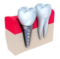
Osteogenesis and morphology of the peri-implant bone facing dental implants Franchi M, Orsini E, Trire A, Quaranta M, Martini D, Piccari GG, Ruggeri A, Ottani V.
This study investigated the influence of different implant surfaces on peri-implant osteogenesis and implant face morphology of peri-implant tissues during the early (2 weeks) and complete healing period (3 months).
Thirty endosseous titanium implants (conic screws) with differently treated surfaces (smooth titanium = SS, titanium plasma sprayed = TPS, sand-blasted zirconium oxide = Zr-SLA) were implanted in femur and tibiae diaphyses of two mongrel sheep. Histological sections of the implants and surrounding tissues obtained by sawing and grinding techniques were observed under light microscopy (LM).
The peri-implant tissues of other samples were mechanically detached from the corresponding implants to be processed for SEM observation.
Two weeks after implantation, we observed osteogenesis (new bone trabeculae) around all implant surfaces only where a gap was present at the host bone-metal interface.
No evident bone deposition was detectable where threads of the screws were in direct contact with the compact host bone.
Distance osteogenesis predominated in SS implants, while around rough surfaces (TPS and Zr-SLA), both distance and contact osteogenesis were present.
At SEM analysis 2 weeks after implantation, the implant face of SS peri-implant tissue showed few, thin, newly formed, bone trabeculae immersed in large, loose, marrow tissue with blood vessels.
Around the TPS screws, the implant face of the peri-implant tissue was rather irregular because of the rougher metal surface.
Zr-SLA screws showed more numerous, newly formed bone trabeculae crossing marrow spaces and also needle-like crystals in bone nodules indicating an active mineralising process. After 3 months, all the screws appeared osseointegrated, being almost completely covered by a compact, mature, newly formed bone.
However, some marrow spaces rich in blood vessels and undifferentiated cells were in contact with the metal surface. By SEM analysis, the implant face of the peri-implant tissue showed different results.
Around the SS screws, the compact bone with areas of different mineralisation rate appeared very smooth, while around the rougher TPS screws, the bone still showed an irregular surface corresponding to the implant macro/microroughness. Around the Zr-SLA screws, a more regular implant-bone surface and sparse, calcified marrow spaces were detectable.
Results from this research suggest that 2 weeks after implantation, trabecular bone represents the calcified healing tissue, which supports the early biological fixation of the implants.
The peri-implant marrow spaces, rich in undifferentiated cells and blood vasculature, observed both 2 weeks and 3 months after surgery, favour the biological turnover of both early and mature peri-implant bone. The implant surface morphology strongly influences the rate and the modality of peri-implant osteogenesis, as do the morphology and arrangement of the implant face in peri-implant bone both during early healing (after 2 weeks) and when the implant is just osseointegrated; rough surfaces, and in particular Zr-SLA, seem to better favour bone deposition on the metal surface.
PMID: 15632988 [PubMed – in process]
Physico-chemical characterization of the surface of 9 dental implants with 3 different surface treatments.
[Article in English, Spanish]
Rodriguez-Rius D, Garcia-Saban FJ. Dr. Daniel Rodriguez, Departamento Cientifico, Impladent S.L, Poligono Industrial Mas d en Cisa, C/ Gato Perez 3-9, 08181 – Sentmenat (Barcelona), Spain.
There are many surface treatments applied to dental implants.
The aim of the present investigation is to compare the physicochemical characteristics of titanium dental implant surfaces with different surface treatments.
9 dental implants from the same batch were divided in 3 groups and received 3 different surface treatments: machined, acid etched and a new chemical surface treatment called Avantblast.
Scanning electron microscopy and confocal microscopy were used to image the treated surfaces, and energy-dispersive spectrometry and X-ray photoelectron spectrometry to provide a chemical characterization of the surfaces.
Results The acid etched and chemical etched surfaces had an increased roughness over the machined one.
Surface chemical composition had differences between processes, as the surface with the new treatment presented a reduced level of impurities and increased thickness of the titanium oxide layer Conclusions Surface roughness of titanium dental implants and thickness of the titanium oxide layer can be increased with a suitable surface treatment.
PMID: 15627909 [PubMed – in process]


השאירו תגובה
רוצה להצטרף לדיון?תרגישו חופשי לתרום!