מתוך medicontext.co.il
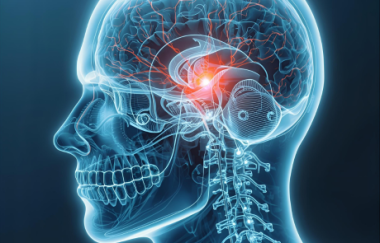
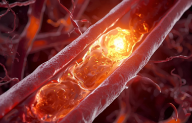
NEW YORK (Reuters Health) – Ruptured fibrous caps that cover lipid cores in the carotid artery, which can be identified by high resolution MRI, are associated with a recent history of stroke and transient ischemic attack (TIA). This suggests that using carotid MRI to identify fibrous cap characteristics may be useful in predicting the risk of stroke.
In Circulation: Journal of the American Heart Association for January 15, Dr. Chun Yuan, from the University of Washington, Seattle, and colleagues report the results of a study of 53 consecutive patients who were scheduled for carotid endarterectomy. Forty-nine of the patients were male, and the mean age of the cohort was 71 years.
Twenty-eight patients had a recent history of stroke or TIA, while 25 were asymptomatic. The researchers performed a carotid MRI on each subject and classified the fibrous cap of each carotid plaque, as intact-thick, intact-thin, or ruptured.
Significantly, 70% of patients with ruptured caps were symptomatic, while only 9% of patients with thick caps and 50% of patients with thin caps were symptomatic (p = 0.001), the researchers found.
When Dr. Yuan's team compared patients with intact-thick caps with patients with ruptured caps, they found that patients with ruptured caps were 23 times more likely to have a history of stroke or TIA.
"These early, promising results suggest that in the future it may be possible to use MRI to track the progress of atherosclerosis and to better select patients for surgical intervention," Dr. Yuan said in a journal statement.

תגובות רוצה להצטרף לדיון?
יש להתחבר כדי להגיב.
התחבראין תגובות עדיין. היה הראשון להגיב!